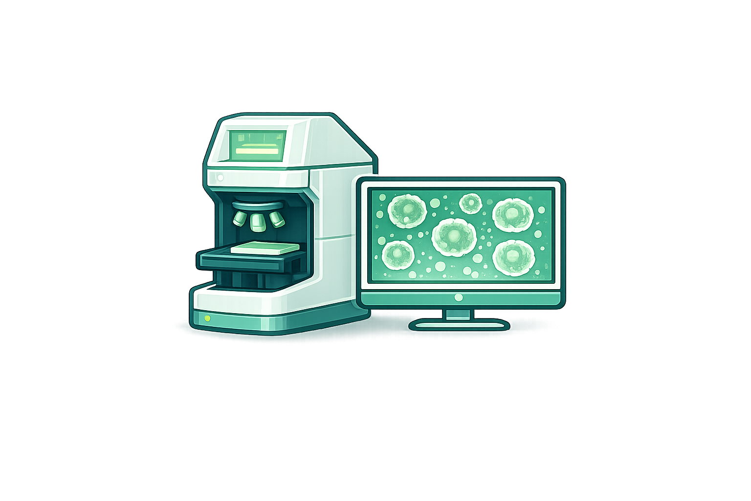
Capture on Your System
Capture brightfield or fluorescence images over time on your existing imaging system.
New application note: Orbits 2.0: Label-free organoid viability that outperforms CellTiter-Glo 3D.
ORGANOID PHENOTYPING PLATFORM
Turn live-cell imaging into quantitative insights for organoids, spheroids, co-cultures, and other complex in vitro model systems.
Faster insights
Automated quantitative readouts from raw imaging.
High-throughput ready
Built to scale across plates, conditions, and timepoints.
Label-free & fluorescence
Analyze multiple modalities in a unified workflow.
Works with your devices
Compatible with the imaging systems already in your lab.
Tailored to your biology
Models optimized for your assays and imaging workflows.
One standardized workflow for any complex in-vitro model — image upload, plate layout, segmentation, quantification, dynamic readouts, and team-wide sharing.
Capture brightfield or fluorescence images over time on your existing imaging system.
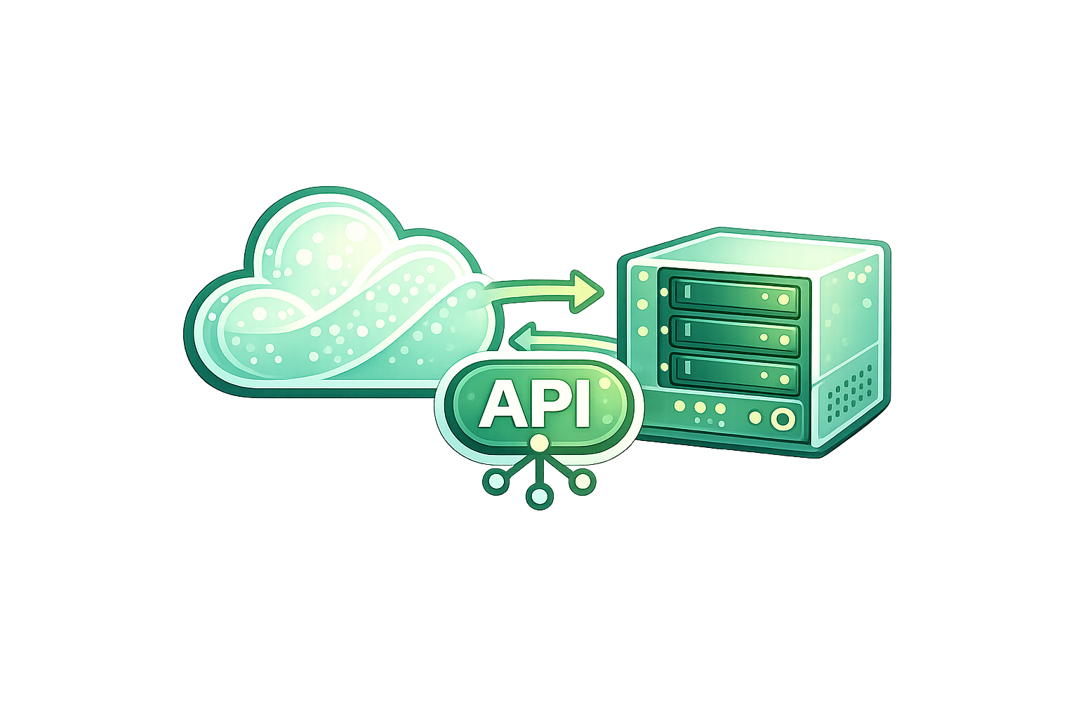
Manage screening experiments across user accounts, with structured images, plate layouts, treatments, timepoints, channels, and replicate metadata.

Detect samples, classify morphologies, and track across the full time course.
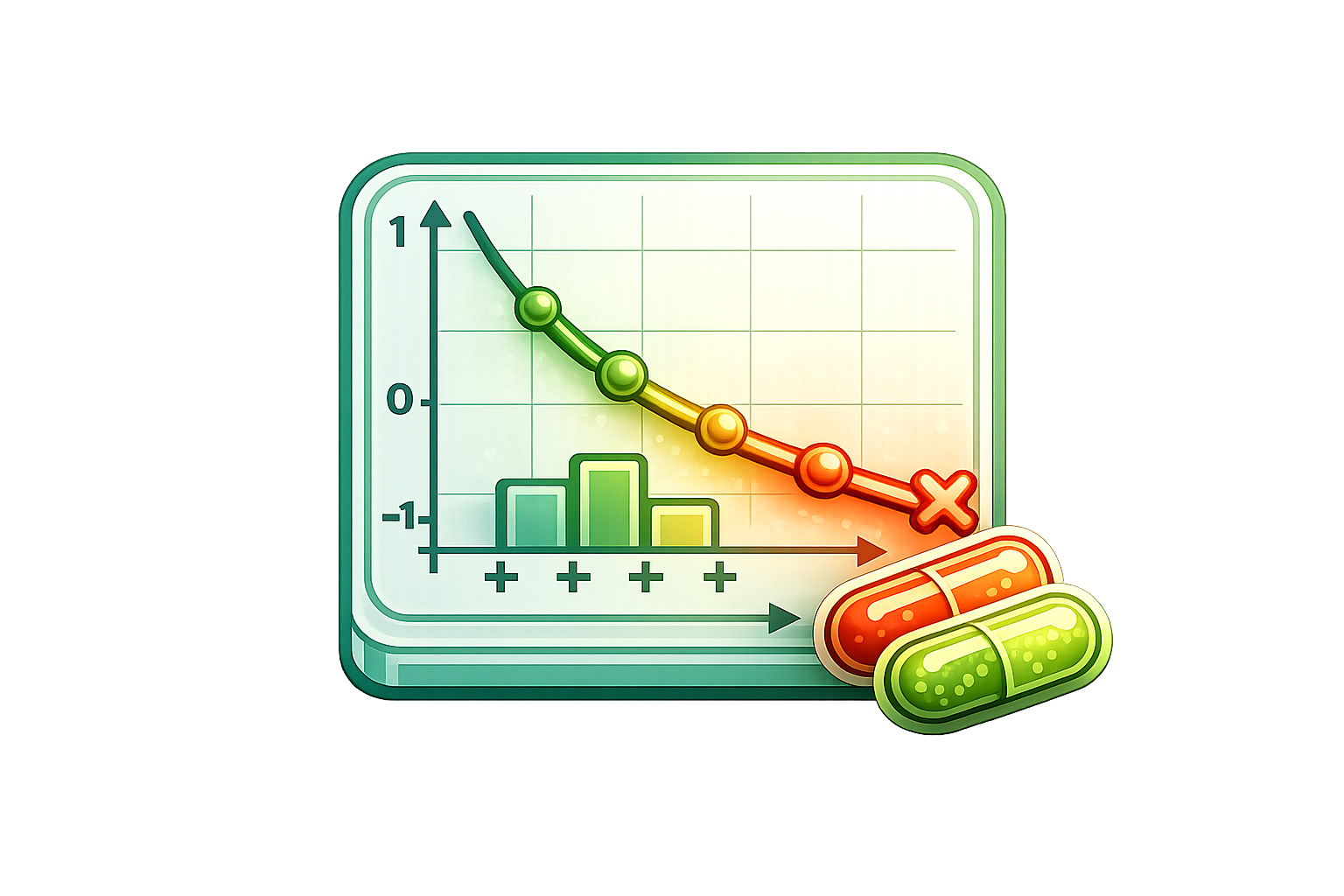
Generate decision-ready metrics from growth, death, morphology, and treatment kinetics.
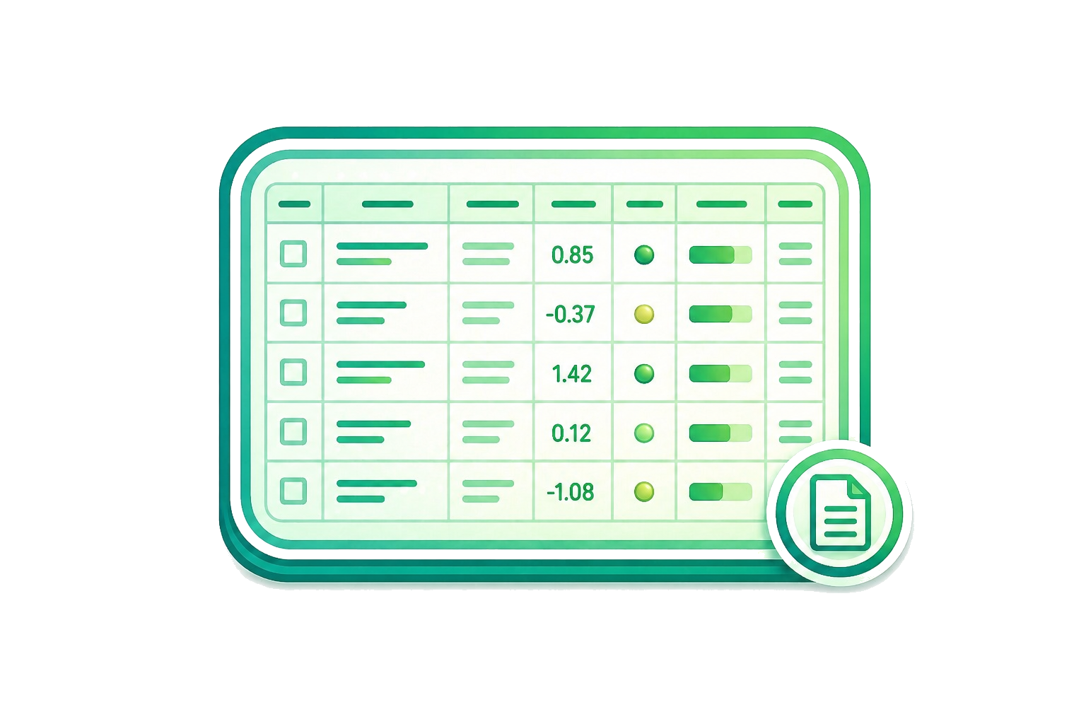
Export analysis-ready results, QC summaries, and structured reports for downstream interpretation.
Device agnostic
Orbits integrates with leading live-cell and high-content imagers for both brightfield and fluorescence workflows.






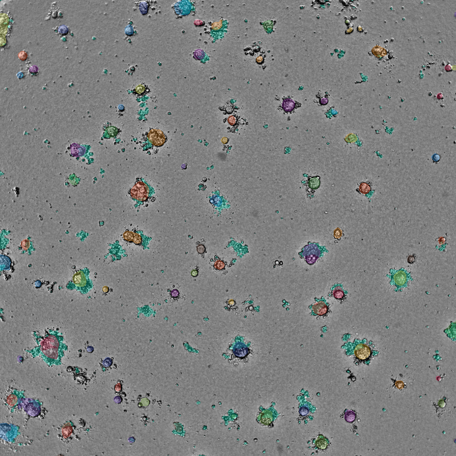
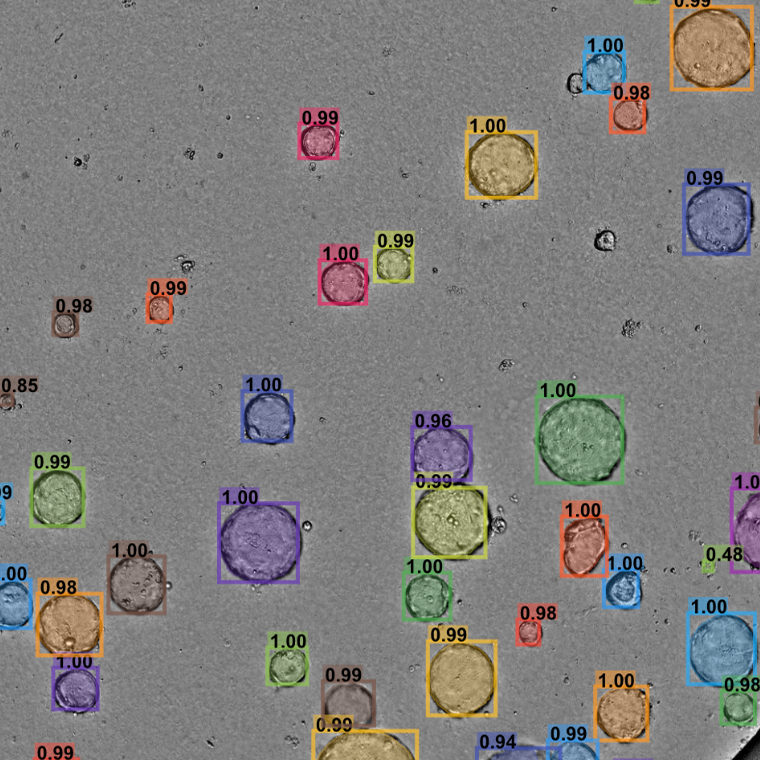
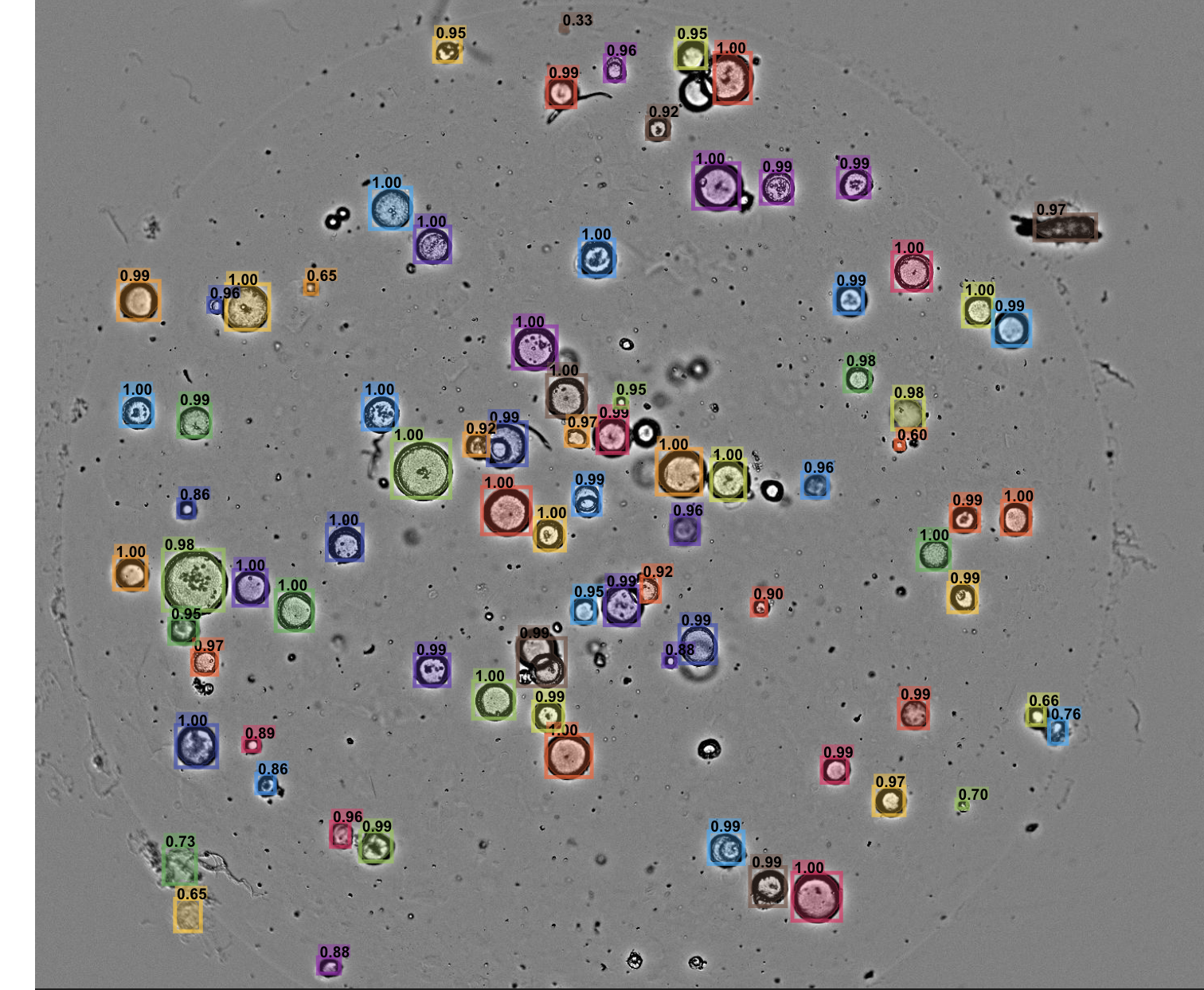
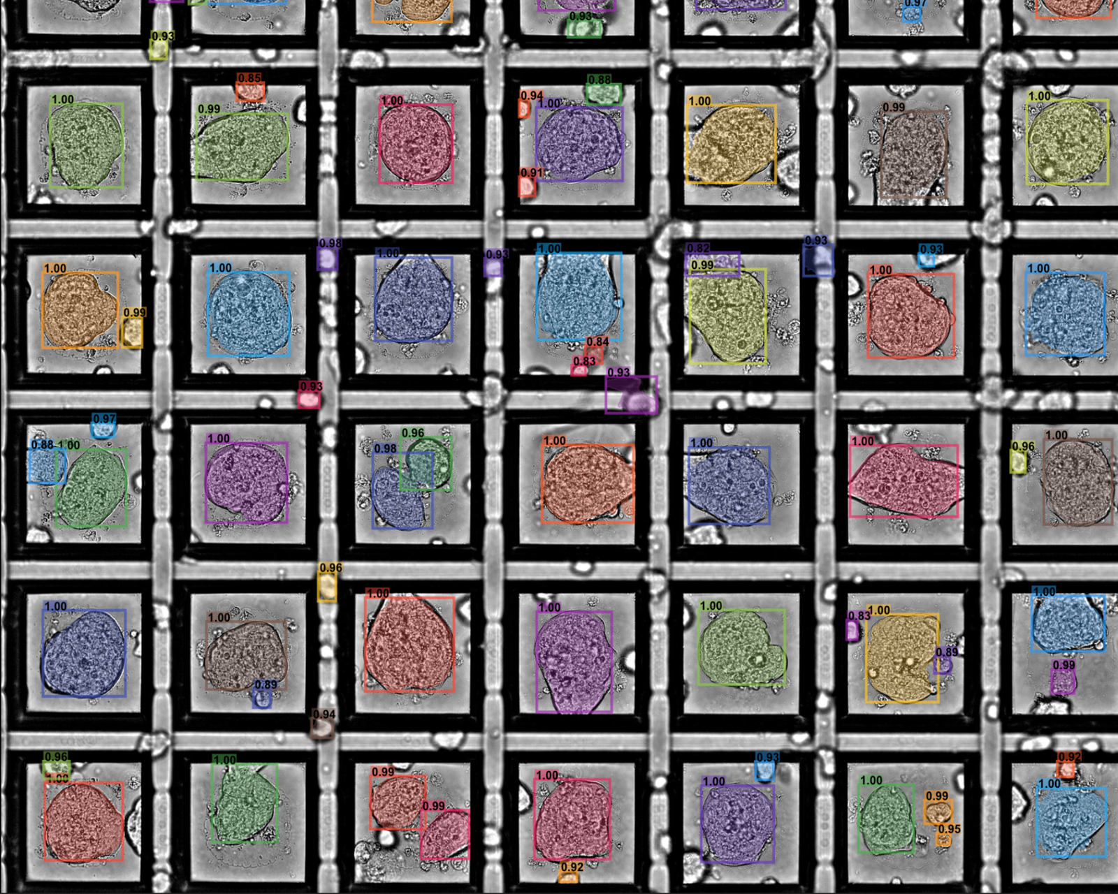
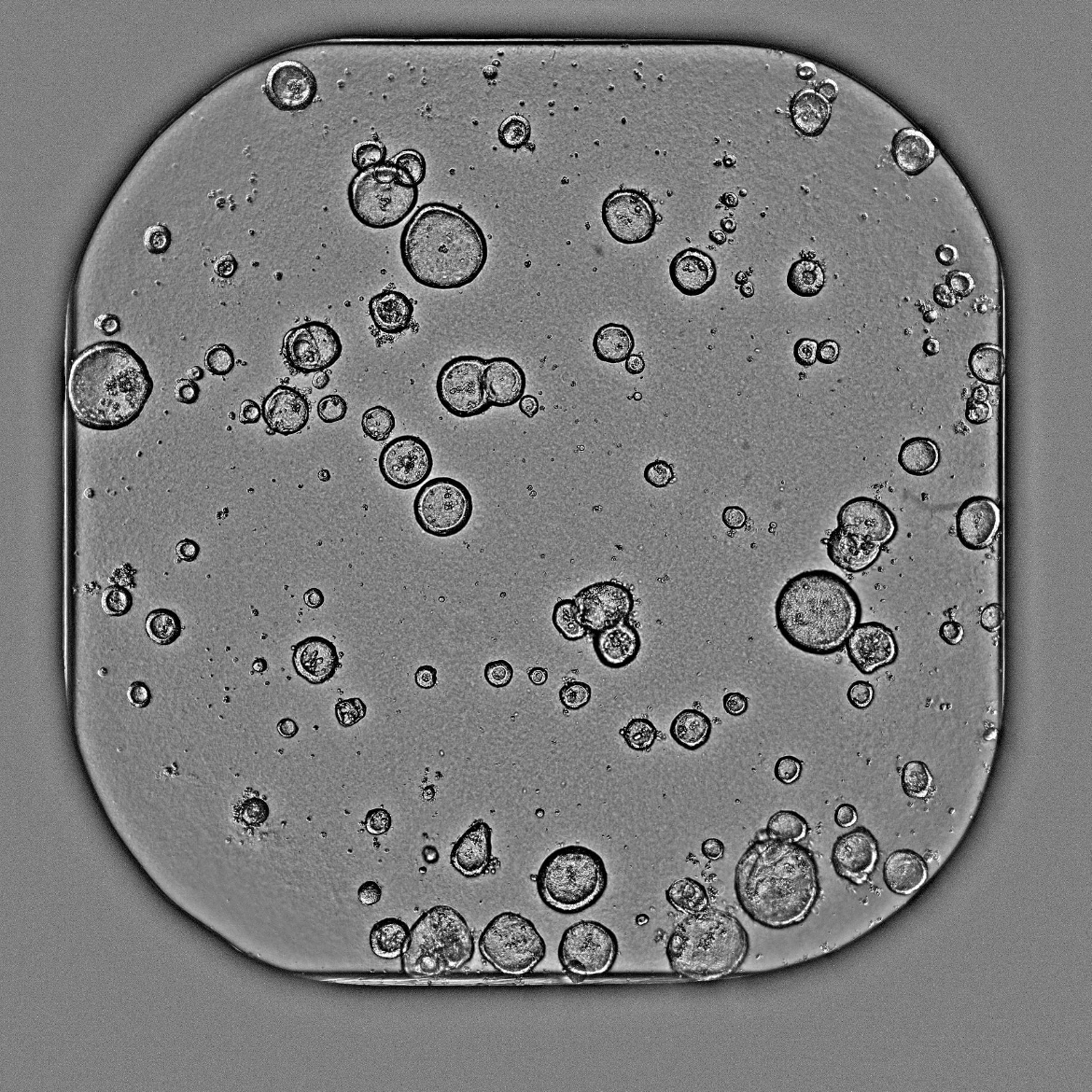
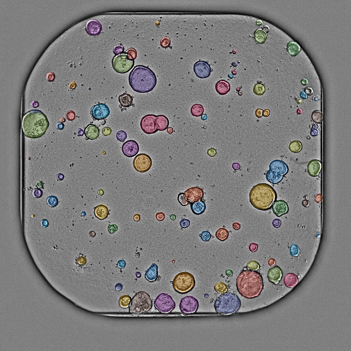
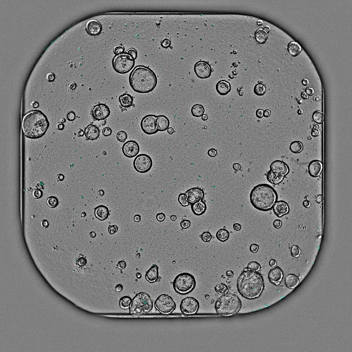
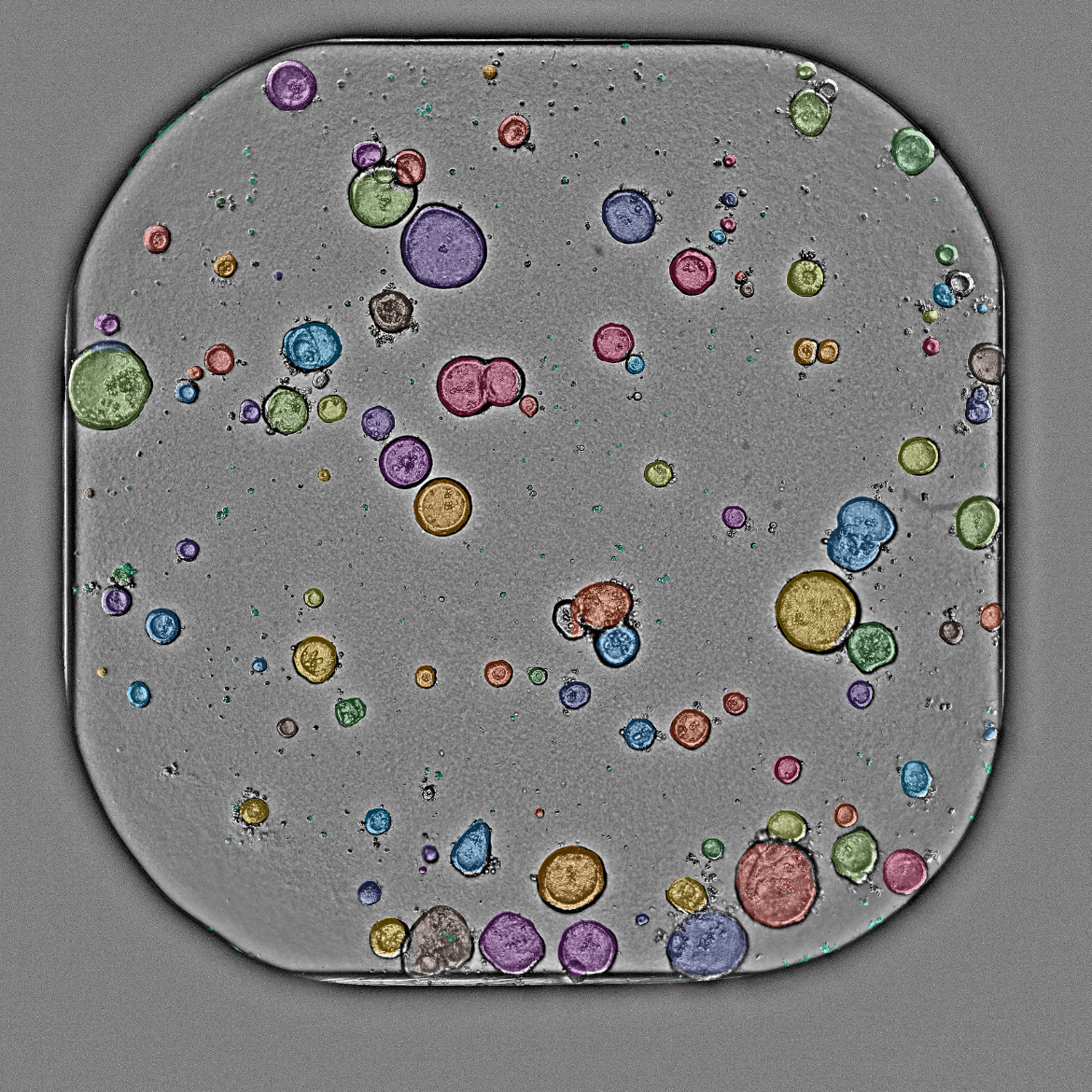
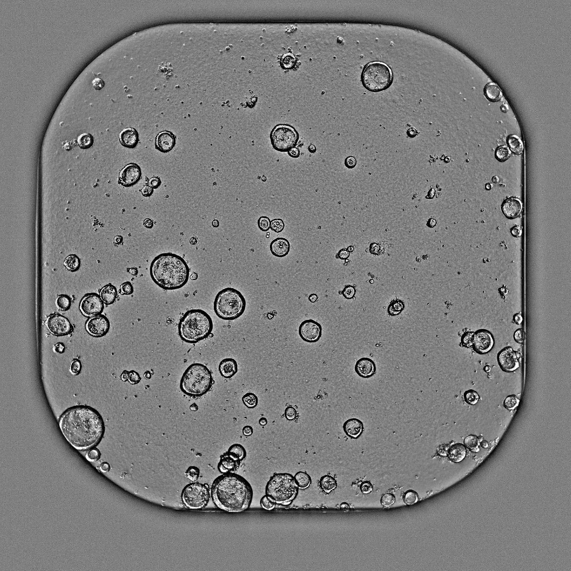
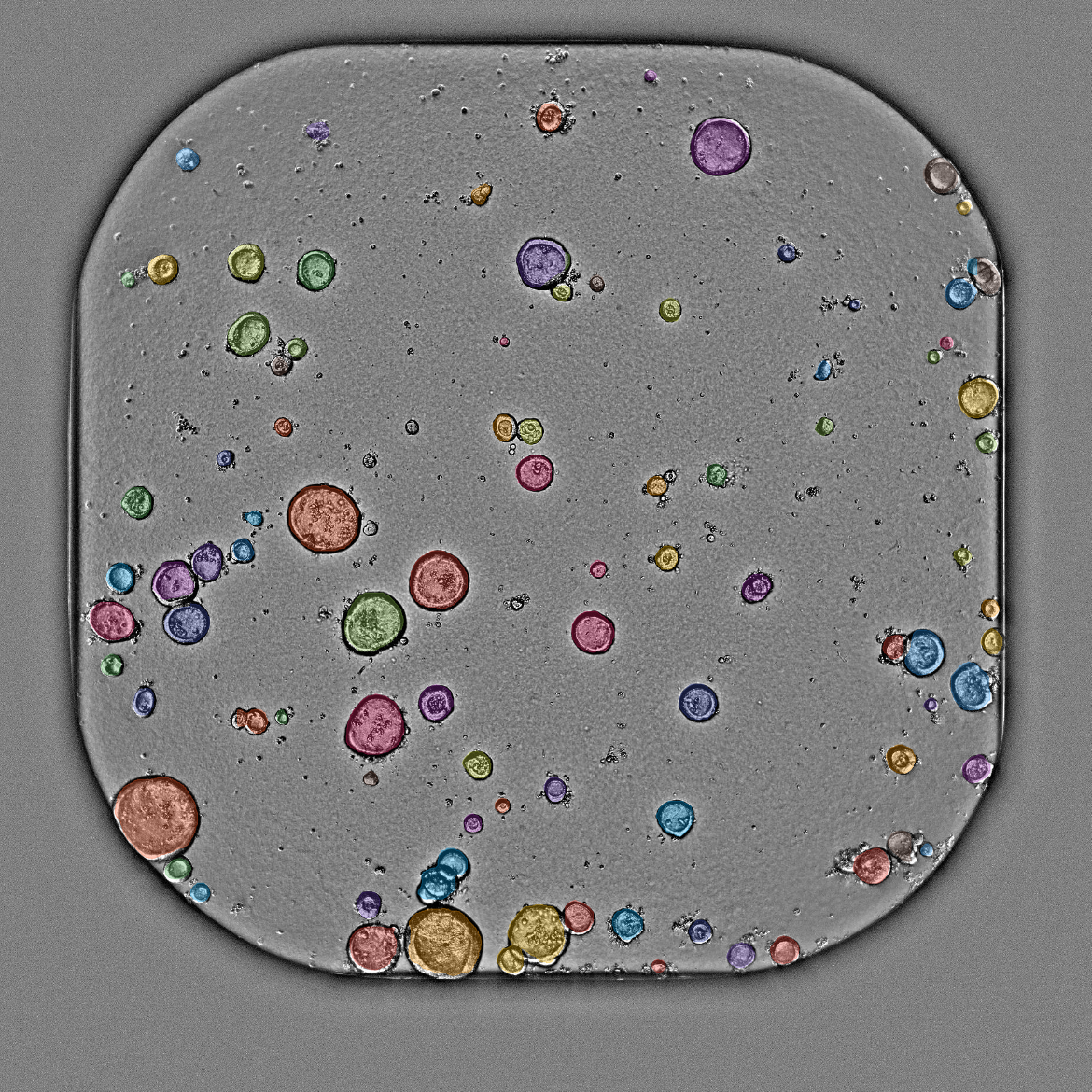
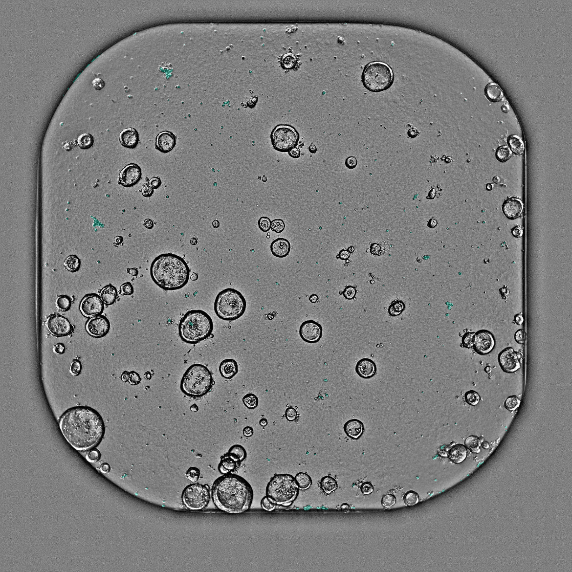
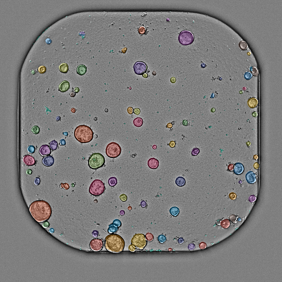
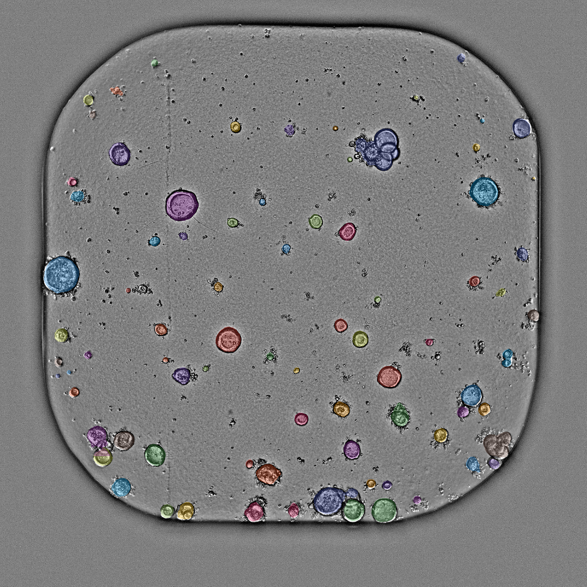
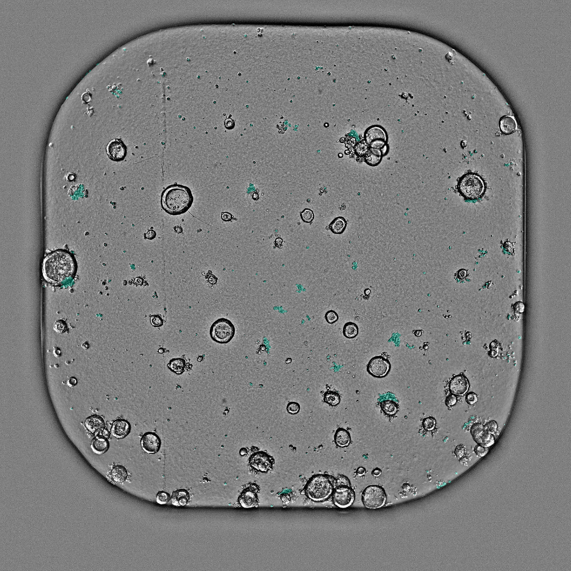
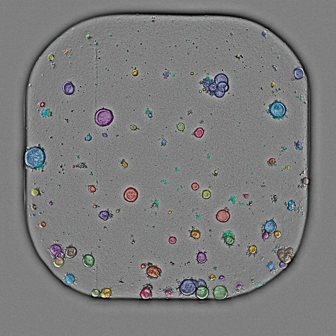
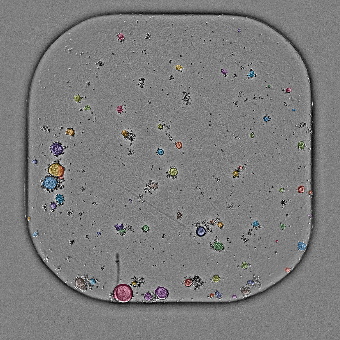
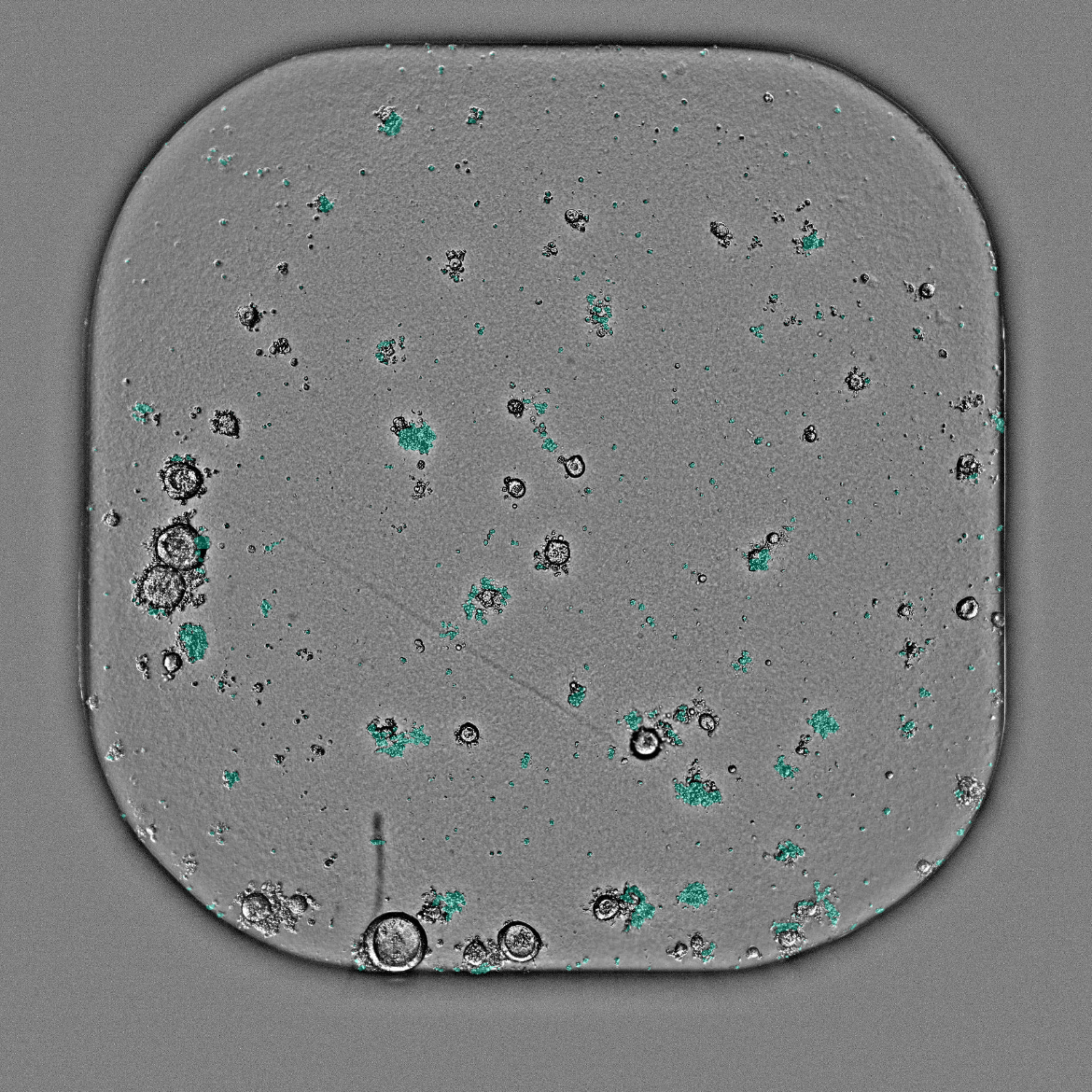
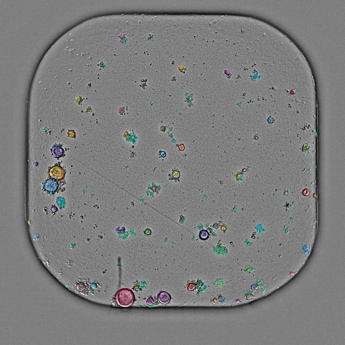
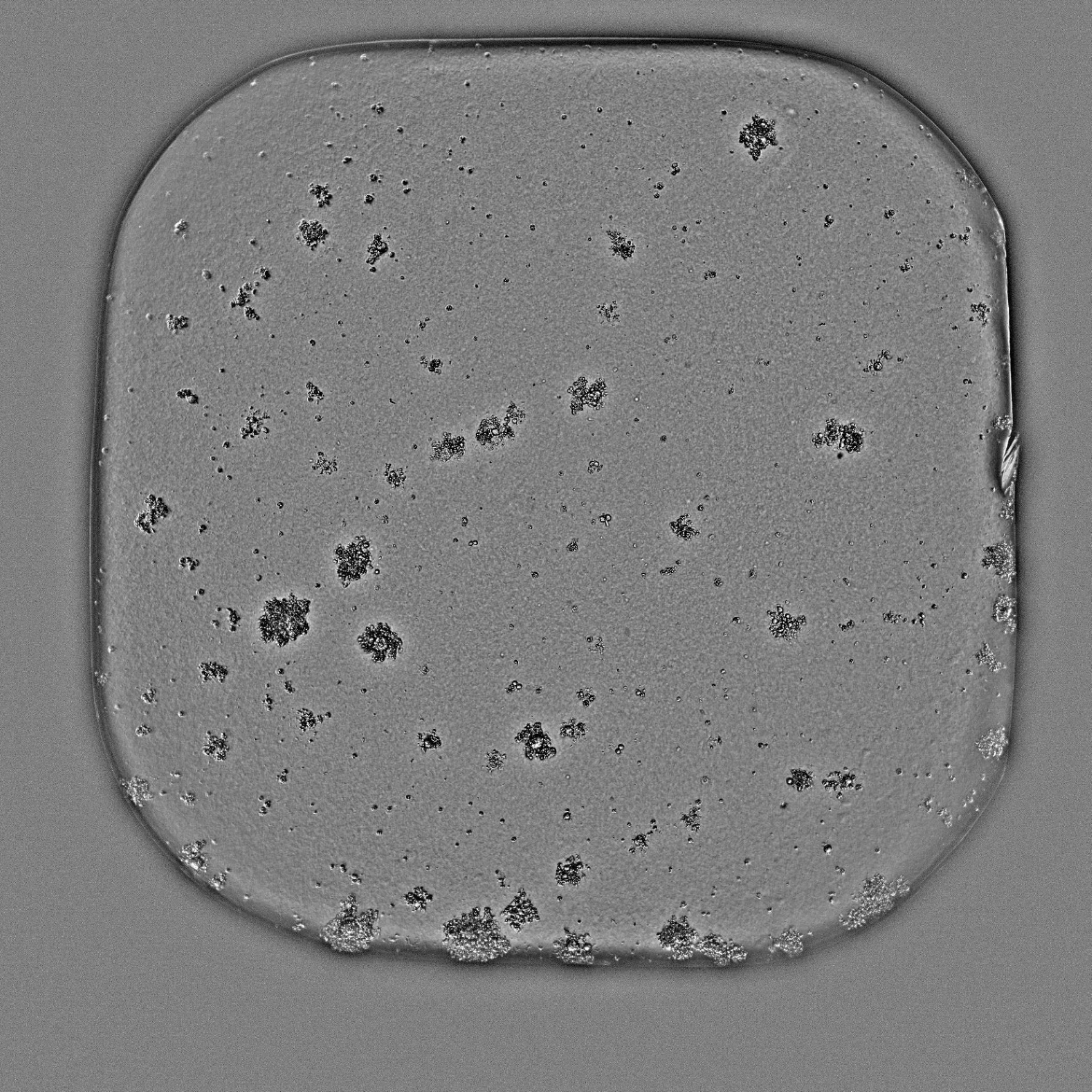
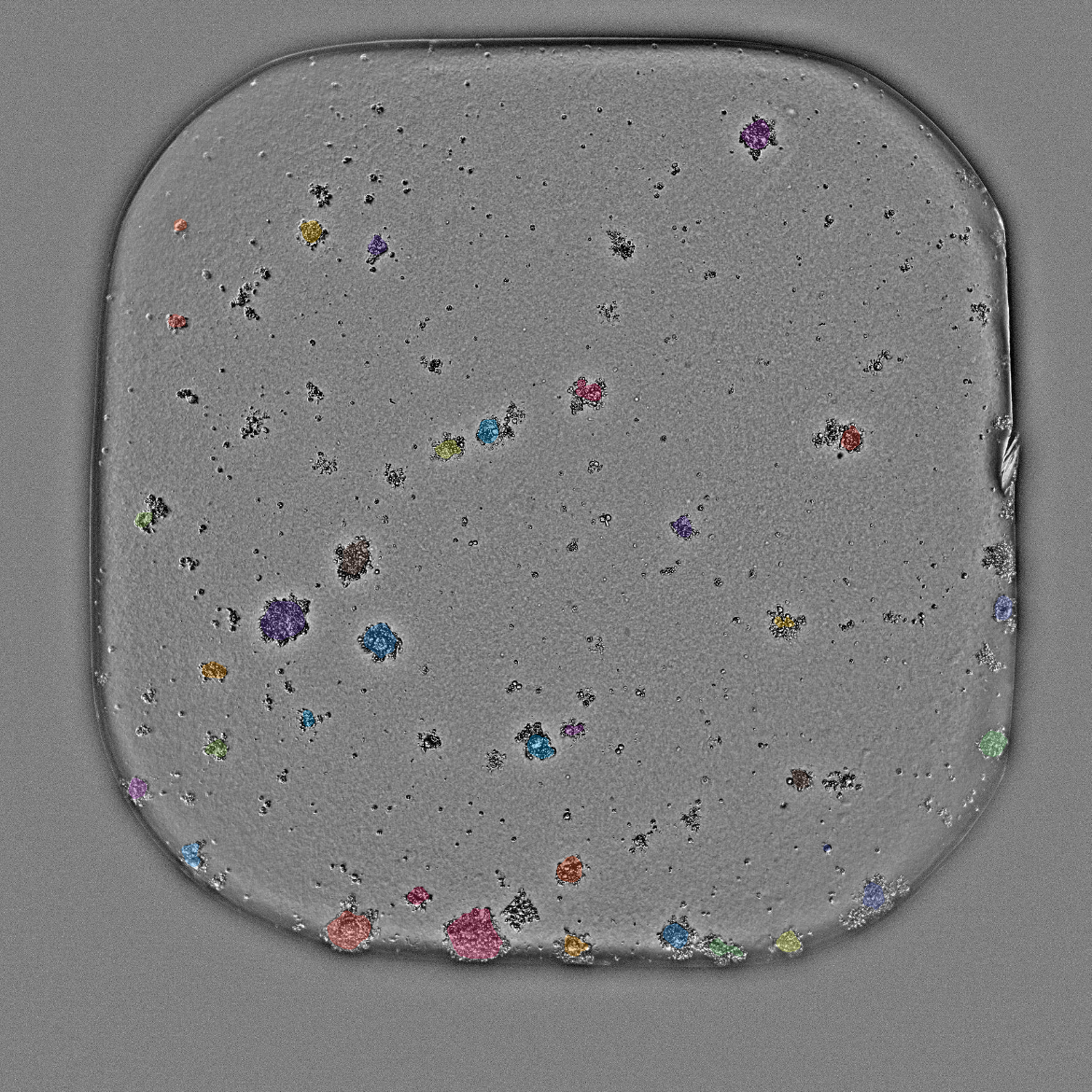
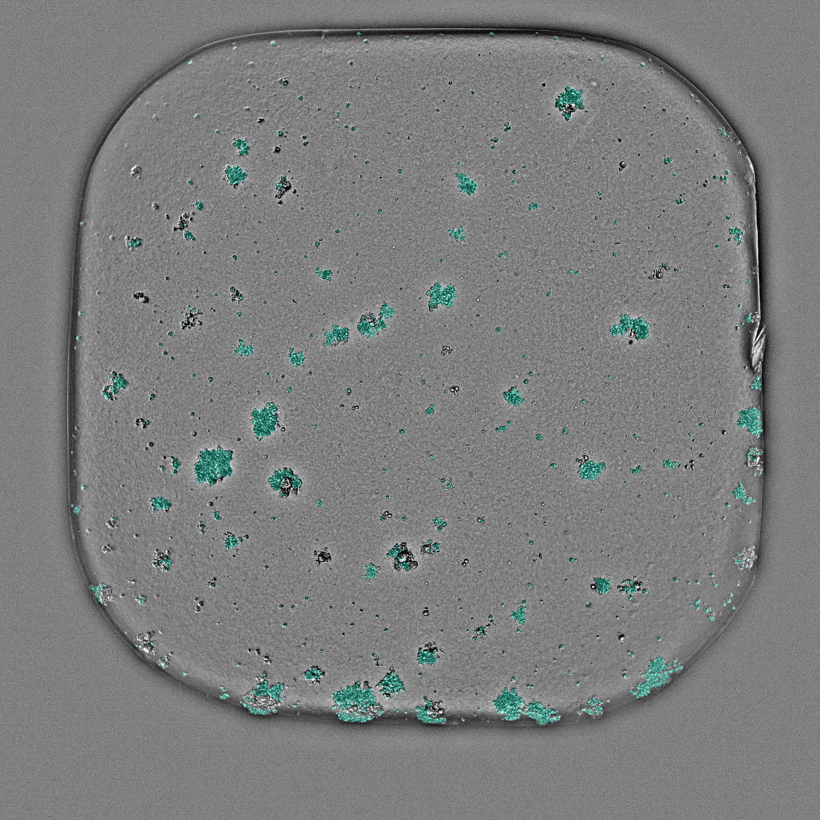
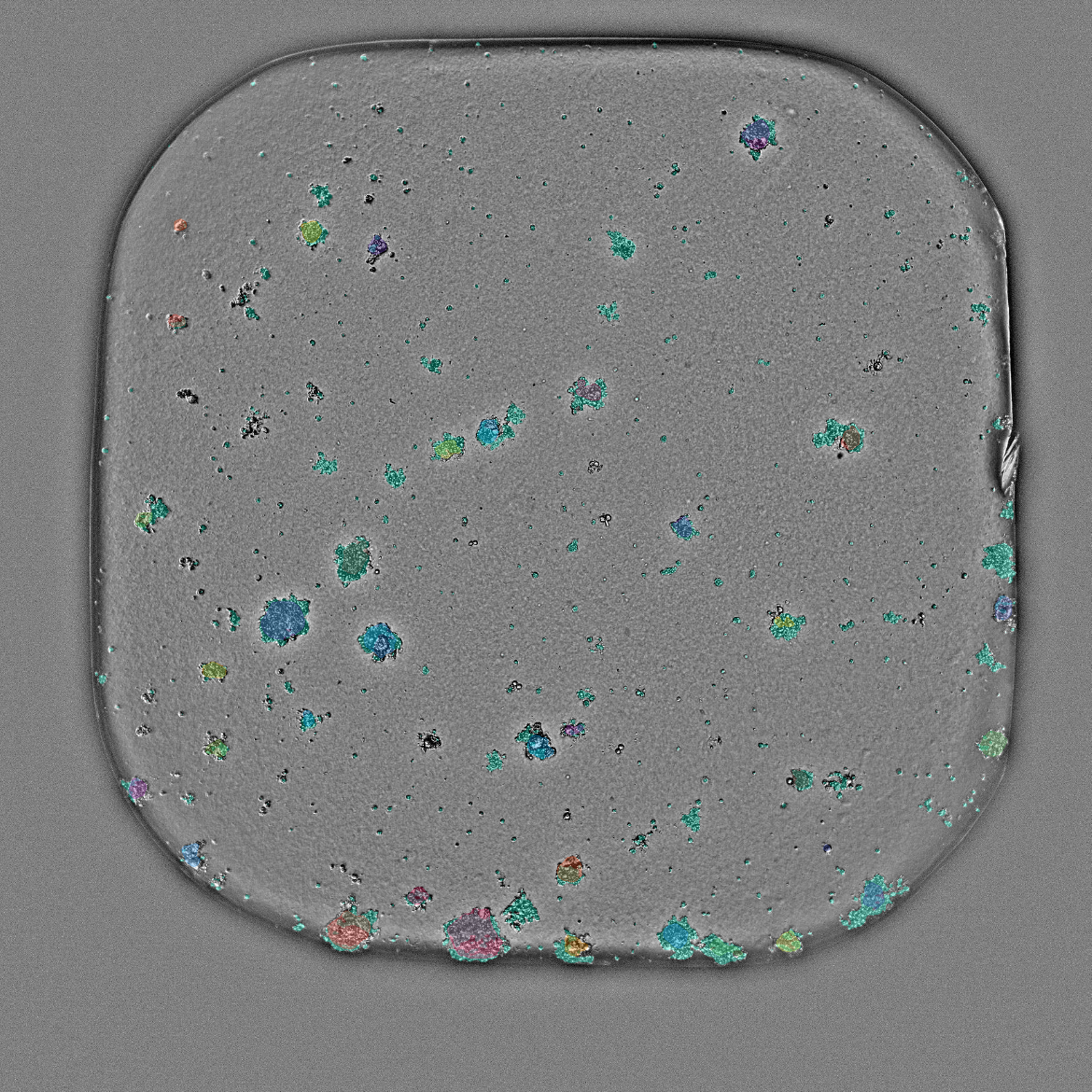
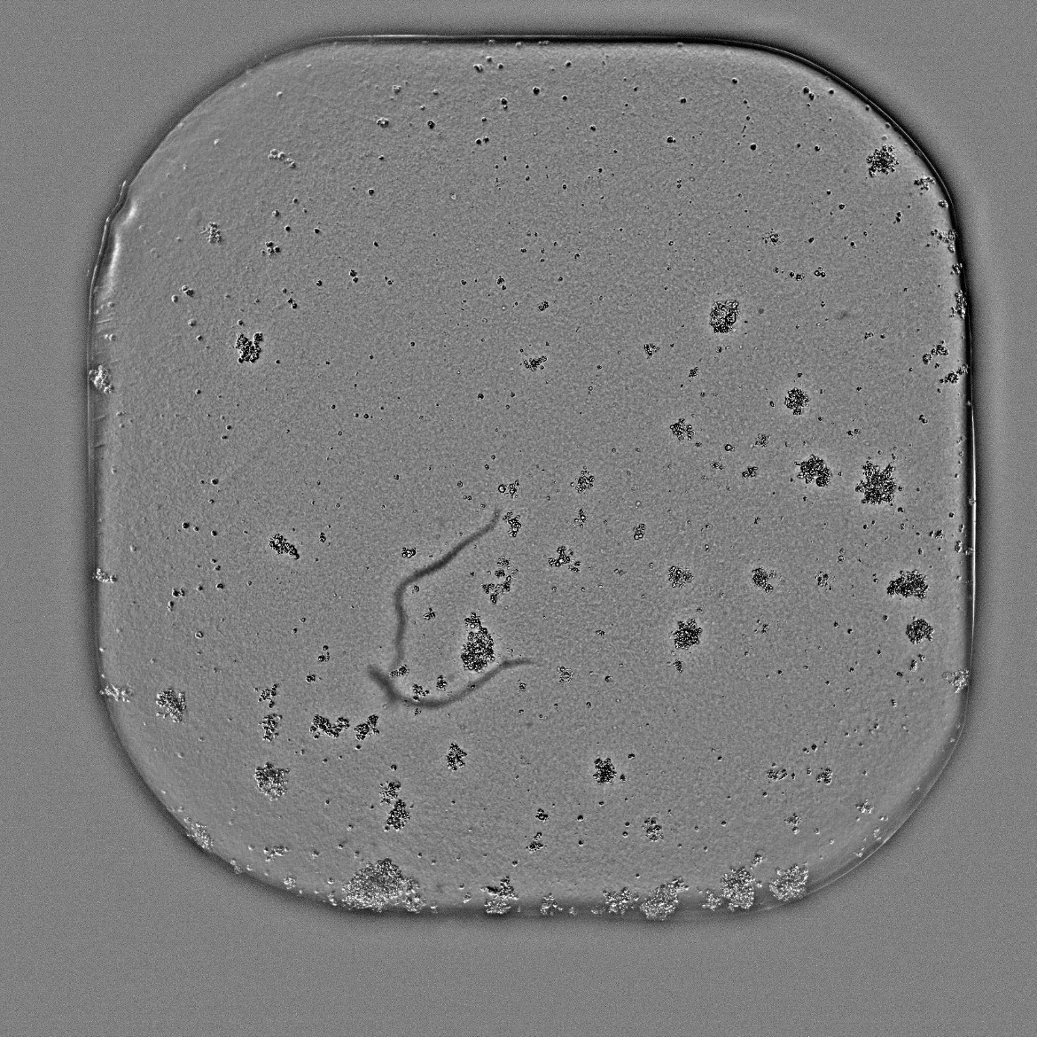
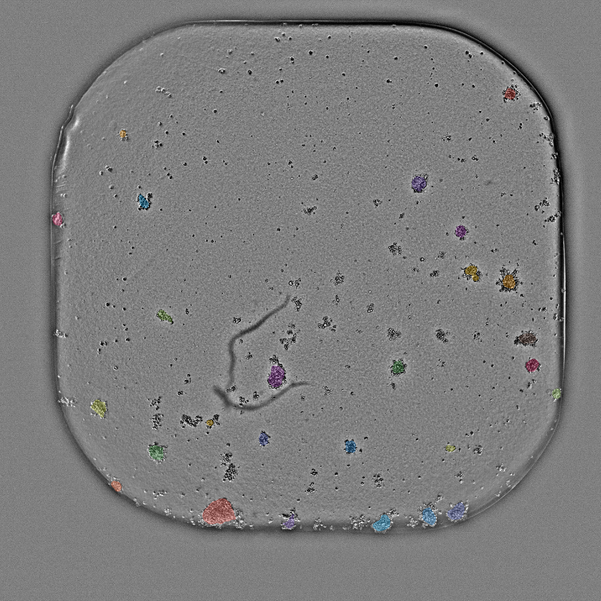
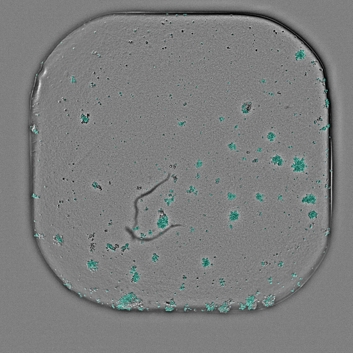
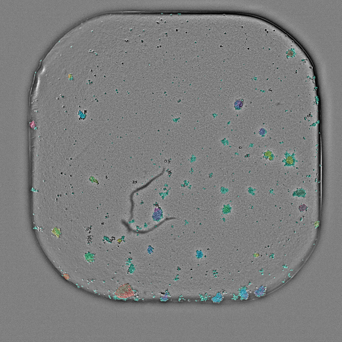
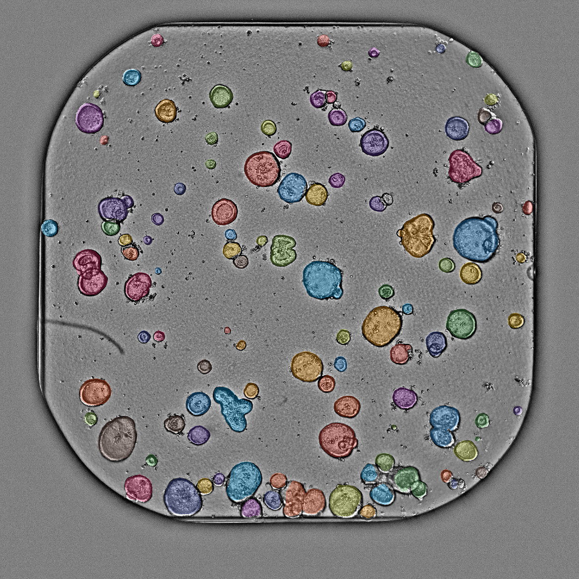
Built from a broad image library of 3D in-vitro models — organoids, spheroids, microtissues, and beyond — Orbits reproducibly segments and quantifies samples, cell death, and treatment-induced phenotypes across diverse morphologies, imaging systems, and plate formats.
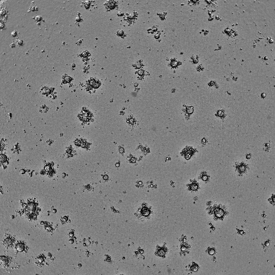
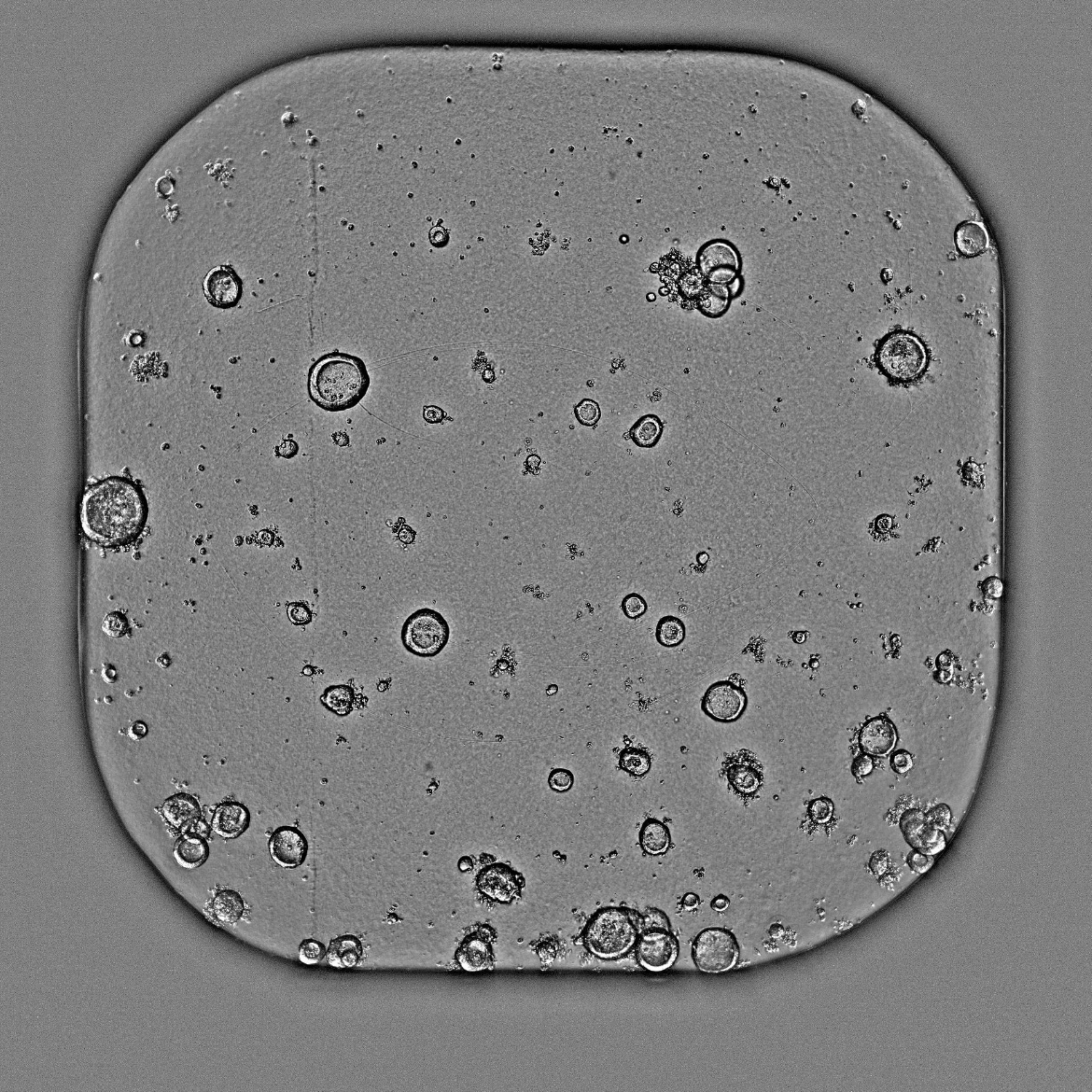
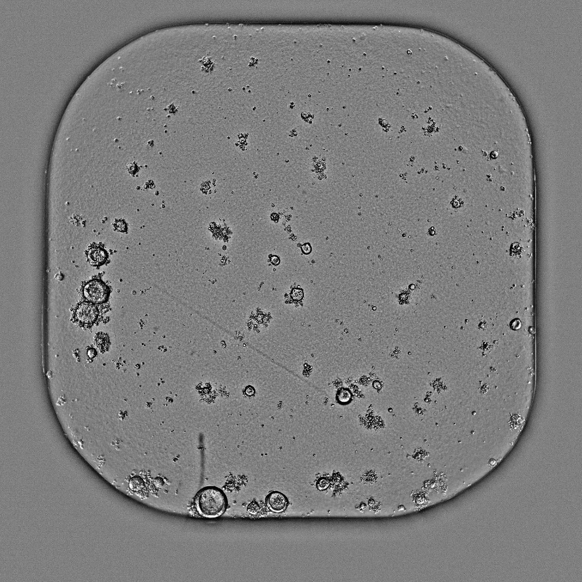
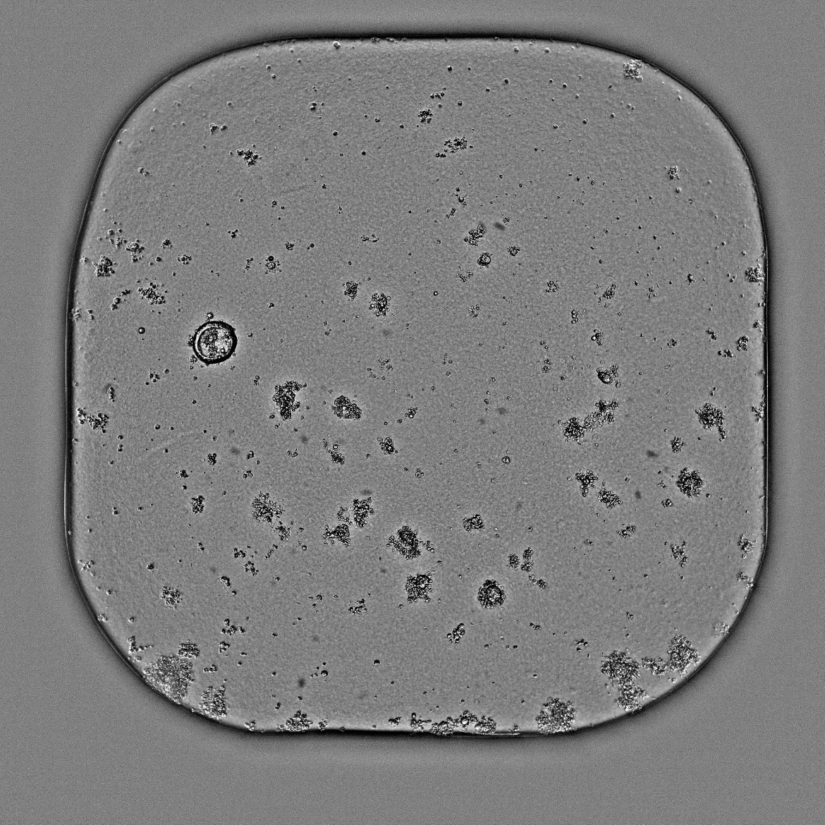
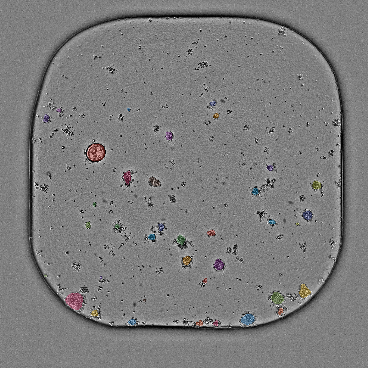
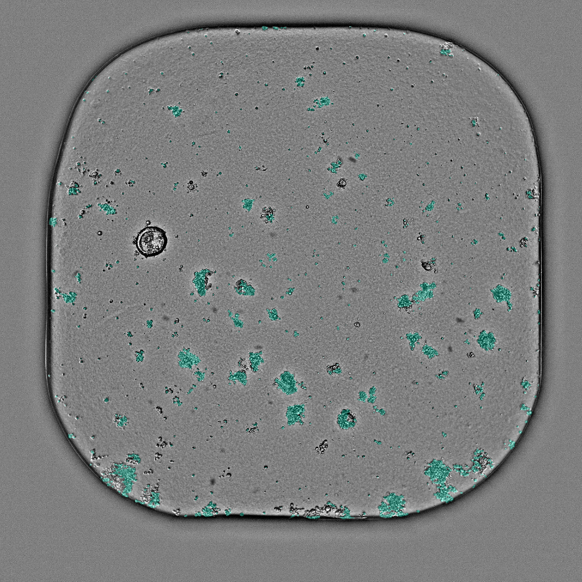
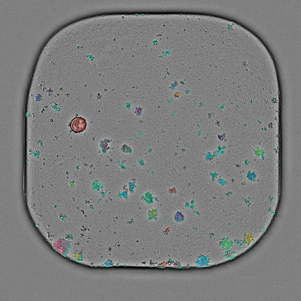
384-well microplate - Corning® #4588 · 4x · Brightfield
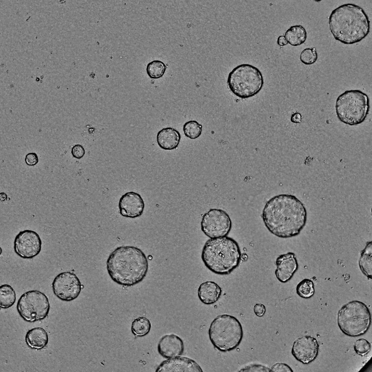
384-well microplate - Corning® #4588 · 4x · Brightfield
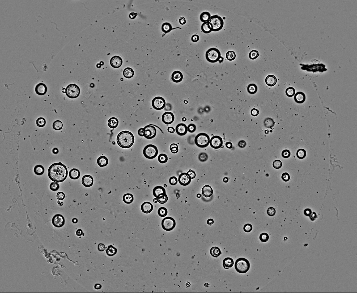
96-well microplate - 4x · Brightfield
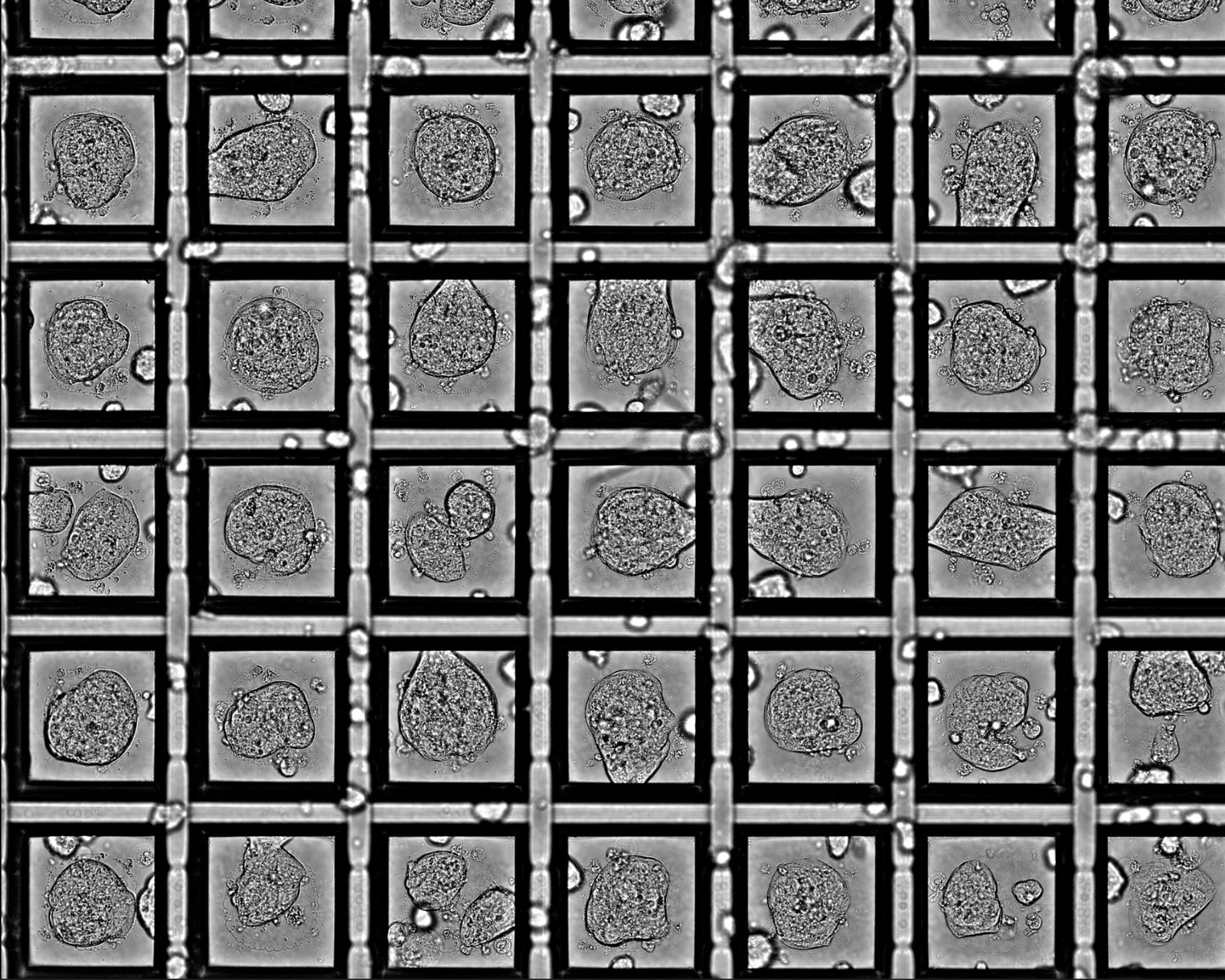
384-well microplate - Corning® Elplasia® #4447 · 10x · Brightfield
From oncology drug screens to toxicology and functional assays, Orbits adapts to the in-vitro models and readouts that matter for your science.
Featured application
Quantify growth rate, morphological heterogeneity, viability, and drug-screening metrics such as IC50, AUC, and our Normalised Organoid Growth Rate (NOGR) for patient-derived tumor organoids. Distinguish cytostatic from cytotoxic responses directly from label-free brightfield images.
Validated head-to-head against CellTiter-Glo 3D in the same wells: r = 0.98 EC50 correlation with 5× lower replicate scatter — no dyes, reagents, or lysis.
Read the application note: Orbits 2.0 vs CellTiter-Glo 3D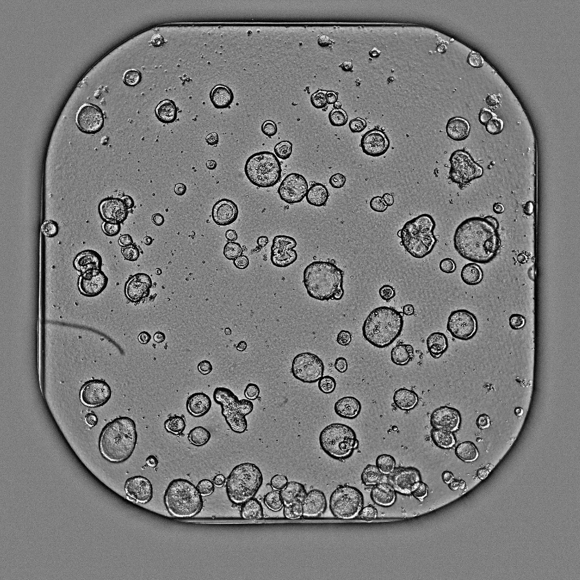
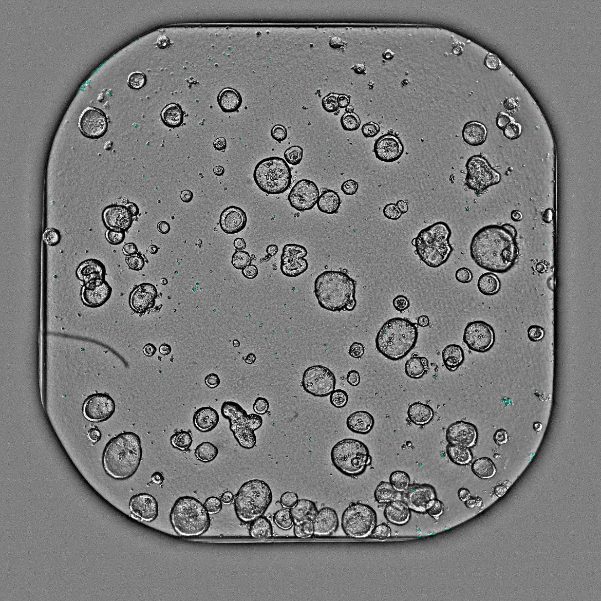
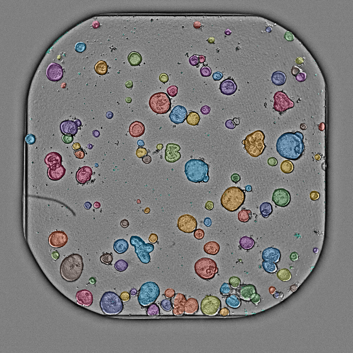
Healthy liver and GI organoids for compound safety and hepatotoxicity screening.
Measure beat rate, amplitude, and contractile dynamics in cardiac organoids.
Morphological and viability readouts for brain organoids and neurospheres.
Working with a different in-vitro model? Let's talk about adapting Orbits to your assay.
Tell us about your in-vitro model and imaging setup. We'll explore whether Orbits can help you get deeper, faster insights from your data.